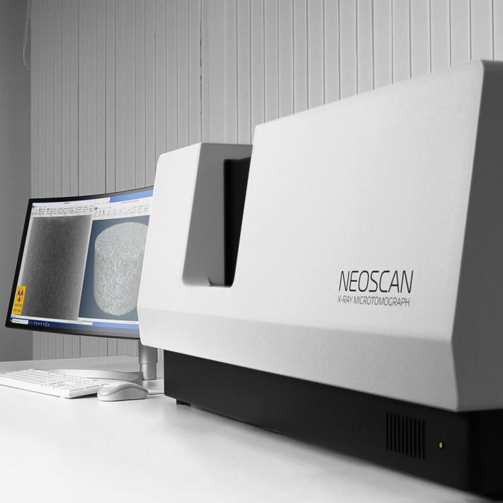
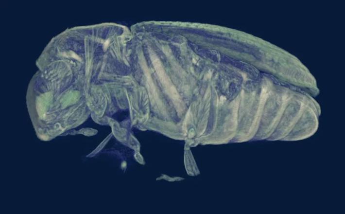
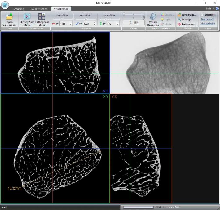
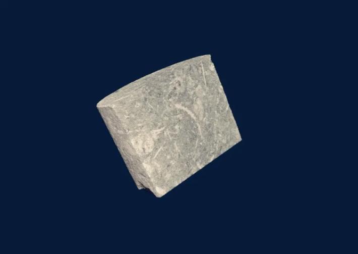
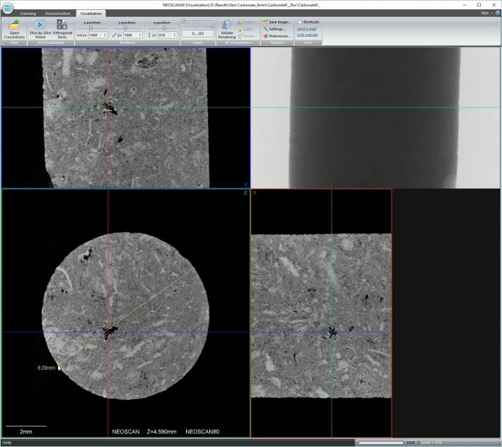
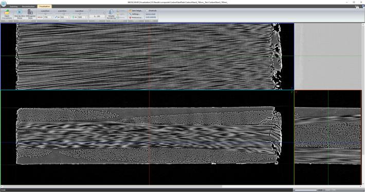
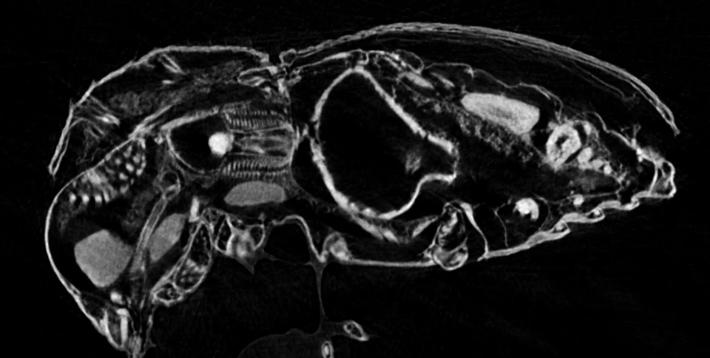
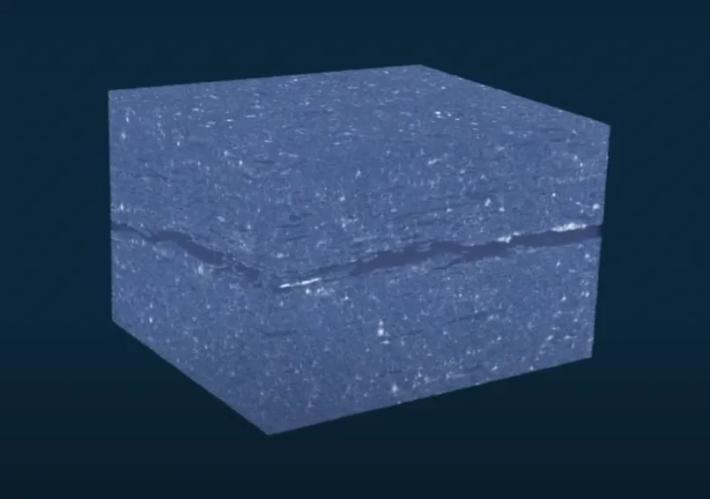
The NeoScan N80 is a scientific grade, high resolution micro-CT scanner, a benchtop, general purpose X-ray laboratory instrument for non-destructive 3-dimensional reconstruction of internal microstructure of the objects with spatial resolution in micron range. Using a X-ray source up to 110 kV this instrument is covering a wide range of applications like geology, composites, electronics, pharmaceutical, bone, dental, and many more. Sub-micron details are detectable due to the phase-contrast enhancement and the low contrast resolution is 2 µm, which is 15x better than microCT instrumentation using a 5 µm spot size.
Videos
News, events, promotions, webinars
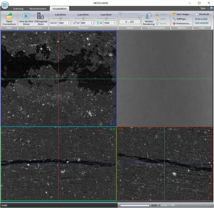
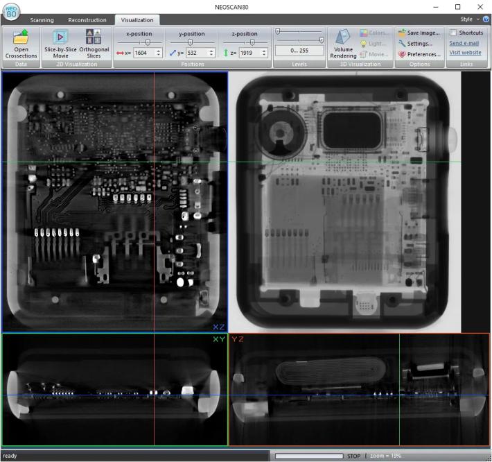
The NEOSCAN N80 is based on newly developed key components, such as microfocus permanently sealed X-ray source and large-format X-ray detector, with specially designed precision electromechanical manipulator, new electronics with micro-stepping drives, and integrated software package for scanning, reconstruction and visualization of obtained results.
The X-ray source has a permanently sealed, maintenance-free construction with unique 2 micrometer spot size, which provides very high spatial resolution of the system. Possibility for target repositioning by operator ensure extremely long lifetime of the source. Using transmission type target allows placing objects very close to emission point and correspondingly reduce distance between the source and X-ray detector with corresponding reduction of scanning time and improvement in signal-to-noise ratio.
The X-ray detector contains large format cooled imaging sensor directly attached to 2:1 fiber-optic taper covered by radiation hardened fiber-optic plate coated by directly deposed scintillator. Such construction with dual fiber-optic design allows 4-fold increase of field of view and ensure strong resistance against radiation damages. Large field of view allows scanning big samples and collecting 4 times more X-ray radiation emitted by X-ray source with corresponding reduction of scanning time and improvement of image quality.
New electromechanical object manipulator in combination with drive electronics for micro-stepping control of all motors provides micron-level accuracy in object positioning and rotation during scanning. The system includes integrated micro-positioning stage on the top of object rotation assembly for exact sample positioning or for selection of scanning volume of interest inside big samples.
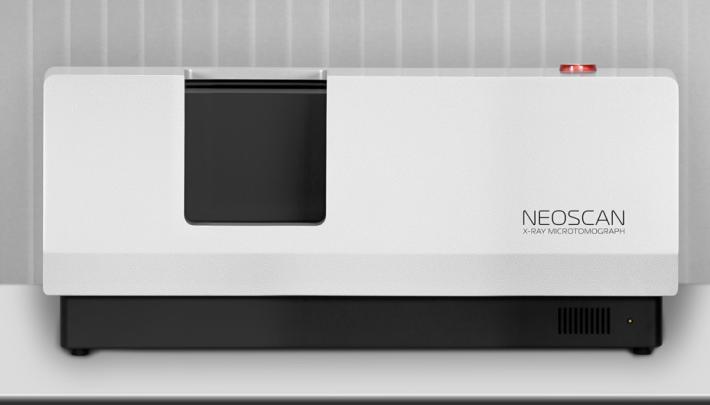
The NEOSCAN N80 system includes internal shielding against radiation leaks sufficient for using this instrument in standard laboratory environment according to all safety regulations without any additional requirements to installation areas. Due to significant weight, the system should be installed on strong, stable desk (not supplied with the system) in vibration-free room.
The system supplied with variety of stages for different object sizes including special stage for large objects with three independently adjusted jaws.
The system can be supplied with optional workstation in one of two standard configurations with all preinstalled software, or with software package on flash drive for installation on customer computer. The system requires one USB2 connection and one FireWire (6-ways) connection to control computer.
Technical Features
Pixel size at maximum magnification | <0.5 um (CCD), <1.2um (FP) |
True low-contrast 3D resolution | 2 um or better |
Maximum scanning diameter (single scan/offset scan)50 mm / 100 mm | 50 mm / 100 mm |
Maximum scanning length | 130 mm |
Maximum physical object length | 200 mm |
Radiation safety | < 1µSv/h at 10cm from instrument surface |
Size / weight | 1200W x 640D x 520H / 225 kg |
Power supply | 100 – 240 V AC, 50 – 60 Hz, 3A |
Shielded feedthrough for user's cables/pipes | yes |
Power & control signals for optional in-situ stages | yes - 24 W (24 V / 1 A) |
Integrated micro-positioning stage | yes - Integrated (10 mm) |
Active artefact suppression | yes |
Type | Maintenance-free, permanently sealed |
Emitter | Transmission target, Tungsten on diamond substrate |
Smallest spot size | 2 µm or smaller |
Maximum voltage | 110 kV |
Maximum power | 16 W |
Automatic filter changer | 20 positions |
Camera type | 7 Mp Flat-Panel or 16 Mp CCD |
Camera field of view (FP / CCD) | 140 x 120 / 72 x 48 mm |
Protection against radiation damage | Yes |
Image sensor | 7 megapixels, cooled |
Camera field of view | 140 x 120 mm |
Scintillator | GADOX |
Fibre-optics | 1:1 |
Protection against radiation damages | radiation hardened fiber optics |